Diagram Of Hip Muscles And Ligaments : Anatomy Of The Hip Joint / The ligaments of the hip joint can be divided into two groups;. A small ligament called ligamentum teres connects the very tip of the femoral head to the acetabular socket. In human anatomy, the muscles of the hip joint are those muscles that cause movement in the hip. It joins the lower limb to the pelvic these ligaments have a unique spiral orientation; Top of the patella and patellar ligament six hip rotator muscles. Muscles allow us to move.
Ligaments are soft tissue structures that connect bones to bones. Ligaments have low vascularity, which means they do not receive much blood flow. The ligaments, tendons, and muscles in the hip joint play a vital role in your ability to walk, run, move, and exercise. A number of ligaments (e.g., iliolumbar ligament, sacroiliac ligament) stabilize and support the joints of the pelvis. A small opening in the muscles and connective tissues of the abdomen — known as the superficial inguinal ring — is located just superior to the inguinal ligament.
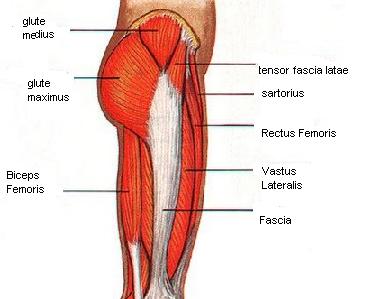
Hip Muscles Pictures And Exercises from www.pilates-back-joint-exercise.com These two muscles produce lateral rotation at the hip and are innervated by the obturator internus and quadratus femoris nerves. Supercial • origin • origin: Tight muscles, tendons, ligaments, and tissues occur with osteoarthritis further limiting joint movement. Joint capsule and ligaments of the hip joint. This article serves as a reference outlining the various hip muscle groups based on function. A number of ligaments (e.g., iliolumbar ligament, sacroiliac ligament) stabilize and support the joints of the pelvis. Muscles creating the movements of the hip joint. • extension of hip • external rotation of the hip.
Ligaments have low vascularity, which means they do not receive much blood flow.
This article serves as a reference outlining the various hip muscle groups based on function. Muscles allow us to move. Equine diagram of tendons and ligaments. Capsular ligaments are intrinsic ligaments of the joint capsule.there are three capsular ligaments that play a key role in maintaining the integrity of the joint during various movements. Tight muscles, tendons, ligaments, and tissues occur with osteoarthritis further limiting joint movement. Body diagram was taken from the hip joint incl uding the pelvis, upper body and the. The foramina of the pelvis (e.g., sciatic foramina, vascular and muscular lacuna) allow the passage of nerves, muscles, blood vessels, and lymphatics. In addition to ligaments, bags are also importantof the hip joint. In addition, the muscles and ligaments. Knee assessment and hip mechanics learn how hip and pelvis mechanics can influence the knee powered by physiopedia start course. Ligaments of the spine provide stability while allowing flexion, extension, and rotation. Ligaments are soft tissue structures that connect bones to bones. Diagram of hip muscles and ligaments move your left leg back up until the top of your thigh rests on the ground.
In human anatomy, the muscles of the hip joint are those muscles that cause movement in the hip. Supercial • origin • origin: Muscles creating the movements of the hip joint. In addition to ligaments, bags are also importantof the hip joint. Muscles allow us to move.
Muscle On Hip Bone from pnpfitness.com This article serves as a reference outlining the various hip muscle groups based on function. Hip joint capsular ligaments serve a fundamental role in balancing functional mobility and joint stability. This causes them to become tighter when the joint is extended. A small ligament called ligamentum teres connects the very tip of the femoral head to the acetabular socket. Equine diagram of tendons and ligaments. Diagram of hip muscles and ligaments move your left leg back up until the top of your thigh rests on the ground. Iliofemoral ligament is the most formidable ligament of body and prevents the trunk from falling backwards in the standing position. Ligaments, tendons, and muscles play an important role in the function of the hip.
We hope this post inspired you and help you what you are looking for.
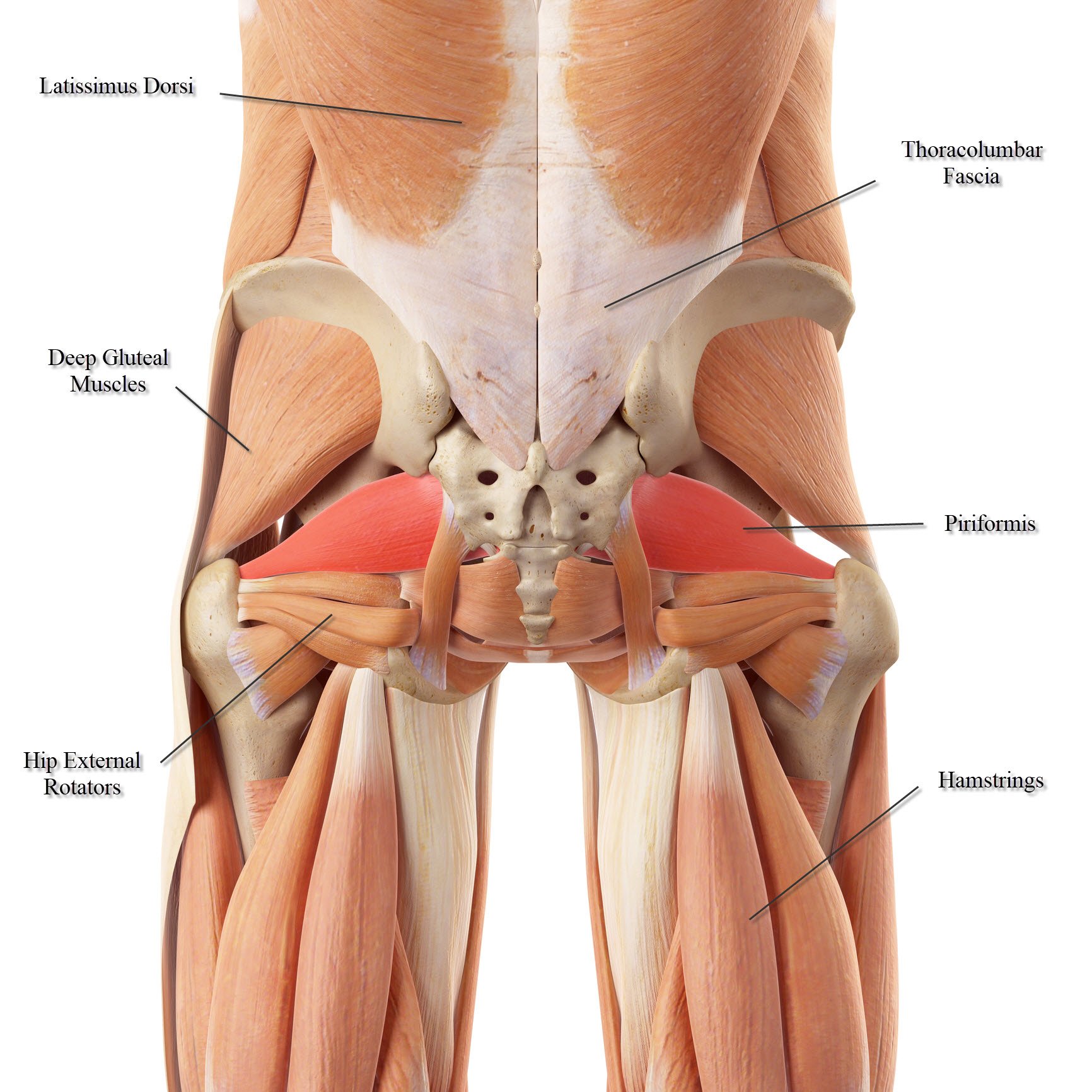
• common action is external rotation • powerful external rotation of the hip is. Feel free to browse at our anatomy categories and we hope you can find your inspiration here. The foramina of the pelvis (e.g., sciatic foramina, vascular and muscular lacuna) allow the passage of nerves, muscles, blood vessels, and lymphatics. Diagram labelled of the hip muscles. There are many different muscles and ligaments in the ankle that may be affected by strains and sprains. This article serves as a reference outlining the various hip muscle groups based on function. Related online courses on physioplus. The inguinal ligament supports the muscles that run inferior to its fibers, including the iliopsoas and pectineus muscles of the hip. It accommodates a small artery within itself that brings an important blood supply to part of the femoral head. Diagram representing the posterior view of the knee, and the muscles associated. A small opening in the muscles and connective tissues of the abdomen — known as the superficial inguinal ring — is located just superior to the inguinal ligament. Ligaments are flexible bands that serve to connect two or more bones together and help stabilize joints. Joint capsule and ligaments of the hip joint.
Ligaments are the structures that bind bones together. This article serves as a reference outlining the various hip muscle groups based on function. Hip joint capsular ligaments serve a fundamental role in balancing functional mobility and joint stability. Ligaments of the spine provide stability while allowing flexion, extension, and rotation. Supercial • origin • origin:
Lower Back Muscle Anatomy And Low Back Pain from ix-cdn.b2e5.com Posts tagged diagram of hip muscles and ligaments. Tight muscles, tendons, ligaments, and tissues occur with osteoarthritis further limiting joint movement. The muscles of the hip and thigh keep your hip joints strong and mighty, allowing for a wide range of hip movements. It joins the lower limb to the pelvic these ligaments have a unique spiral orientation; Knee assessment and hip mechanics learn how hip and pelvis mechanics can influence the knee powered by physiopedia start course. It accommodates a small artery within itself that brings an important blood supply to part of the femoral head. One or more ligaments provide stability to a joint during rest and movement. A small ligament called ligamentum teres connects the very tip of the femoral head to the acetabular socket.
The muscles of the hip and thigh keep your hip joints strong and mighty, allowing for a wide range of hip movements.
Thank you for visiting muscles and ligaments of the hip pictures. We hope this post inspired you and help you what you are looking for. Tight muscles, tendons, ligaments, and tissues occur with osteoarthritis further limiting joint movement. Ligaments are soft tissue structures that connect bones to bones. A number of ligaments (e.g., iliolumbar ligament, sacroiliac ligament) stabilize and support the joints of the pelvis. It joins the lower limb to the pelvic these ligaments have a unique spiral orientation; Capsular ligaments and intracapsular ligaments. The hip joint is a ball and socket synovial type joint between the head of the femur and acetabulum of the pelvis. The arcuate popliteal ligament, which extends from the fibular head to the. Related online courses on physioplus. Accessory anterior inferior tibiofibular ligament. A small ligament called ligamentum teres connects the very tip of the femoral head to the acetabular socket. Muscles creating the movements of the hip joint.
This lack of blood flow makes ligaments slower to heal than other types of soft tissue hip muscles diagram. Supercial • origin • origin:


0 Komentar